

Planning radiotherapy with PET/CT raises treatment effectiveness and reduces complications
One of the latest innovations in radiotherapy is being applied in Venezuela. PET/CT reveals metabolic lesions that a conventional scan cannot show, say doctors Luisa Suárez and Giuseppe Figurelli.
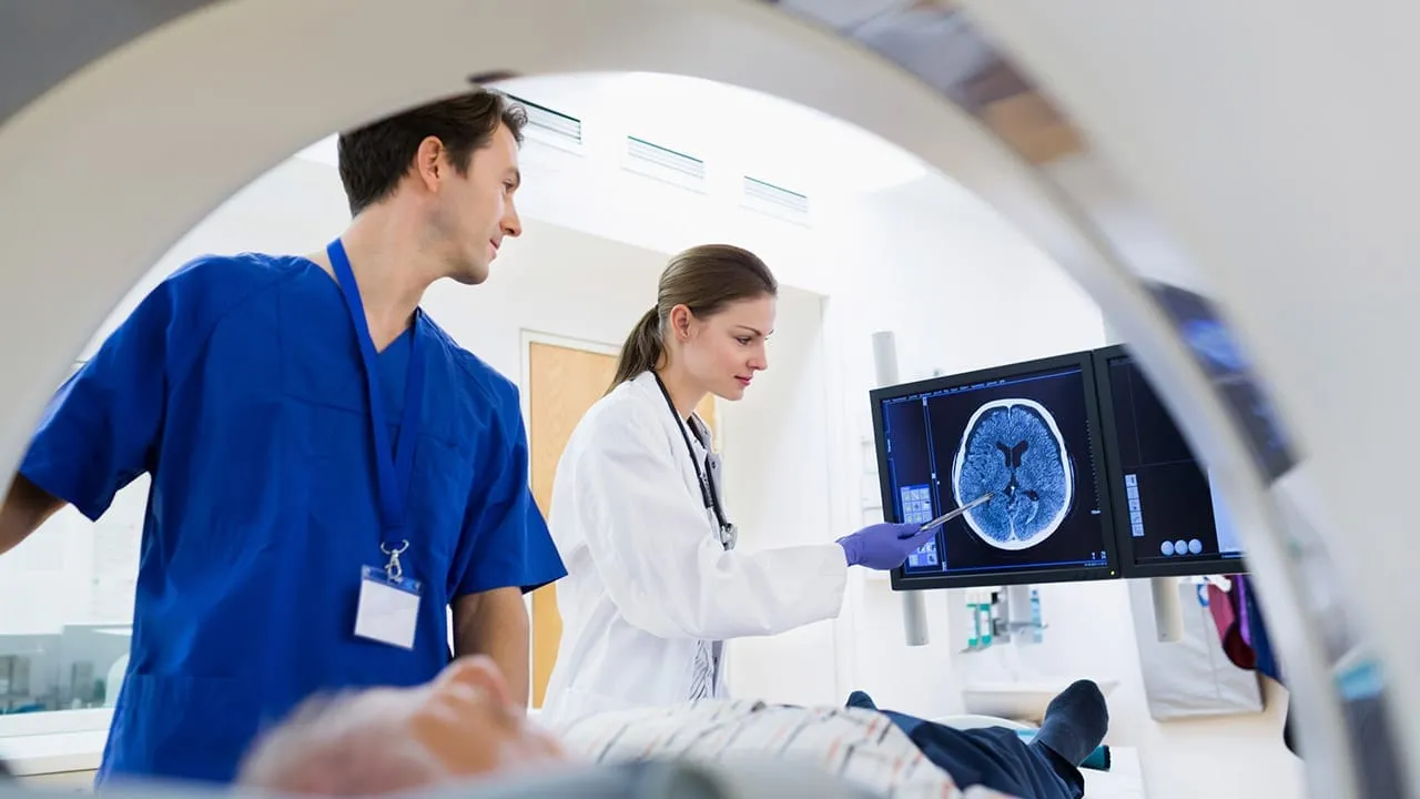
Scientists keep working to reduce cancer mortality through treatment techniques that avoid complications for patients and make the experience of recovery as bearable as possible.
Venezuela is applying the technique of planning radiotherapy with PET/CT, a study that allows clinicians not only to see the patient's anatomy statically but also to distinguish how any lesion behaves metabolically. Metabolic lesions are not visible in a conventional scan, but PET/CT can see beyond that frontier, making it possible to reduce or increase the volume treated with radiotherapy and avoiding over or under dosing in treatment.
Cancer is one of the leading causes of death worldwide. According to the World Health Organization, around 16 per cent of the world's population dies of the disease, on 2015 figures, while in Venezuela 2019 closed with approximately 28,000 deaths, according to the study Forecast of Cancer Mortality and Incidence in Venezuela, produced by the Venezuelan Anti Cancer Society with Universidad Simón Bolívar.
Luisa Suárez and Giuseppe Figurelli are doctors who graduated from Universidad Central de Venezuela and specialise in oncological radiotherapy at Centro Diagnóstico Docente Las Mercedes, an institution leading in medical technology in Venezuela and Latin America, directed by Wilson Mourad, a specialist in the digestive system.
Forecast of cancer mortality and incidence in Venezuela
From the story
Suárez and Figurelli said the fundamental purpose of applying radiotherapy planning with PET/CT is better dosing for patients, since the Achilles heel of radiotherapy is that the beam has to pass through healthy tissue, and if the entry dose or the volume to be irradiated can be reduced, complications are avoided.
"You can also increase the radiation dose to the volume you want to irradiate, and that is achieved with PET/CT, in which hybrid images are fused to give better conformation of the radiation, with a higher dose in tumour tissue and a lower one in healthy tissue. In other words, there is better discrimination between what will be irradiated and what will not," the specialists said.
In the view of Suárez and Figurelli, it is ideal for lymphomas and for lung, rectal, gastrointestinal, head and neck and prostate tumours, since these are tumours that can be visualised with the different PET markers, FDG and PSMA, helping to delimit them more accurately, reducing the tissue to be irradiated or allowing reductions that lead to higher doses under international protocols.
They added that PET/CT should be performed on all patients for staging, saving time that is extremely valuable for oncology patients, since a single examination avoids other studies such as scans, ultrasounds and scintigraphy. It also defines the area to be irradiated after surgery in cancers that require it.
PET/CT is a great ally in planning radiotherapy and can be performed on all patients for whom the study is indicated. Every tumour has its treatment protocol. We know which are operable and which are not, and we delimit volumes accordingly. "PET/CT becomes a tool that helps the radiation oncologist achieve greater accuracy when planning and delivering radiation in patients for whom radiotherapy and chemotherapy are used for the radical cure of the disease," Suárez and Figurelli said.
Follow this story every morning
The NotiCorp newsletter, free and with no paywall.